Novel Tissue Harmonic Imaging Clearly Visualizes a Case of Intraductal Papillary Mucinous Neoplasm with Mural Nodules
DOI:
https://doi.org/10.6092/1590-8577/2385Keywords:
Diagnostic Imaging, Endosonography, Pancreatic NeoplasmsAbstract
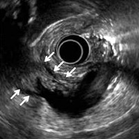
Tissue Harmonic Echo (THE) imaging is a sonographic technique that potentially provides images of higher quality than can conventional B-mode images. Potential advantages of THE imaging include improved resolution, improved signal-to-noise ratio, and reduced artifacts [1, 2]. Recently, a novel THE imaging performed using an EUS system with a monitor/processing unit (EU-ME2 PREMIER PLUS; Olympus Medical Systems, Tokyo, Japan) has been developed. Using this technology, we can obtain two THE mode images, namely, THE-P (penetration) and THE-R (resolution). The THE-P mode is suitable for middle range distance observation because it receives a harmonic signal whose frequency is mainly 7.5 MHz. The THE-R mode is suitable for close distance observation from the probe because it receives a harmonic signal whose frequency mainly ranges from 10 to 12 MHz. Here, we report a case of intraductal papillary mucinous neoplasm (IPMN) with mural nodules which could be clearly detected using this novel THE imaging.
Image: P-mode image.
Downloads
References
Shapiro RS, Wagreich J, Rarsons RB, Stancato-Pasik A, Yeh HC, Lao R. Tissue harmonic imaging sonography: evaluation of image quality compared with conventional sonography. Am J Roentgenol. 1998; 171:1203-1206. [PMID:9798848]
Ishikawa H, Hirooka Y, Itoh A, Hashimoto S, Okada N, Itoh T et al. A comparison of image quality between tissue harmonic imaging and fundamental imaging with an electronic radial scanning echoendoscope in the diagnosis of pancreatic diseases. Gastrointest Endosc. 2003; 57:931-936. [PMID:12776049]